The new specimen is named Pterygornis dapingfangensis, which is a relatively advanced enantiornithine. An external sternal manubrium develops on the front edge of the sternum. In living birds, the external sternal manubrium is mainly used to attach the clavicular membrane of the pectoralis, but this structure has not been reported in other Early Cretaceous birds.
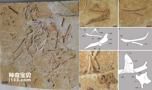
Figure 1 The true form and partial skull skeleton of the Great Bungalow-winged Bird
Although the early Cretaceous enantiornithine specimens are relatively complete, different bones overlap each other, so the morphological characteristics of the skulls are difficult to observe. The bones of the new specimen are scattered, especially the small skull bones, which are well preserved, making it possible to restore part of the shape of the skull, especially the zygomatic bones and square zygomatic bones.
According to researchers, the zygomatic bone and the square zygomatic bone come from different ossification centers. During the embryonic stage of living birds, the two have completely healed into a rod-shaped bone, forming the lower edge of the orbit. In dinosaurs, close relatives of birds, the zygomatic bone and the zygomatic bone do not unite. The posterior end of the zygomatic bone bifurcates to form the postorbital apophysis and the zygomatic zygomatic process. The zygomatic bone is in a "T" shape, pointing forward respectively. , project the zygomatic process, squamous process and ventroposterior process dorsally and posteriorly. In dinosaurs, the postorbital apophysis of the zygomatic bone articulates with the postorbital bone, thereby completely separating the orbit from the inferior temporal fossa; the squamous apophysis of the quadratozygomatic bone articulates with the squamosal bone, forming the closed posterior edge of the inferior temporal fossa. Compared with dinosaurs, the skulls of living birds have undergone tremendous changes, especially the degeneration of the posterior orbital bone, the loss of the zygomatic bone-postorbital bone joint, and the quadratozygomatic bone-squamous bone joint. The changes allow birds to move the square bones in the front-to-back direction to push the bones of the palate forward and backward, ultimately allowing the mouth to be raised or lowered relative to the head to complete feeding activities.
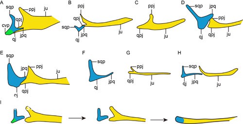
Figure 2 The evolution of square cheekbones and cheekbone shapes in dinosaurs and birds
For a long time, due to the preservation of fossils, it has not been clear how the above-mentioned changes in the bones behind the orbit occurred during the evolution of dinosaurs and birds. Through detailed comparisons, Wang Min et al. discovered for the first time that the square cheekbones of Pterornis are similar to those of Archaeopteryx, Confuciusornis, Huipornis, Jeholornis, and Archaeorhynchus. They all lost the ventral and posterior lateral processes, forming an "L"-shaped skeleton. Compared with dinosaurs, the square zygomatic bone and zygomatic bone became slender in primitive birds, and the squamous apophysis and postorbital apophysis degenerated significantly. It is speculated that the zygomatic bone - postorbital bone and the zygomatic bone - squamous bone joints have been lost in birds of the Early Cretaceous, and the "T"-shaped square zygoma went through an "L"-shaped transition stage during the evolution to a short-stalked shape, and first lost its ventral and posterior sides. process, indicating that this skull feature that is conducive to birds' feeding activities has appeared in the early stages of bird evolution.
animal tags:
We created this article in conjunction with AI technology, then made sure it was fact-checked and edited by a Animals Top editor.
you may also like

Sponges are the simplest structure and the most primitive form among multicellular animals. They have appeared before the Cambrian and have continued to thrive until modern times. Sponges evolved from single-celled animals. Their cells have differentiated but have not yet formed tissues and o...

Kuban pigs (kubanochoeres) are a type of huge pigs with hump-shaped teeth that once lived in the Old World. They were mainly distributed in the Early Miocene to Middle Miocene, and are found in Africa and Eurasia. For a long time, paleontologists have been debating whether the Cuban pig should be es...

Although Anderson noticed as early as 1921 that there were some white bladed vein quartz fragments at the site where the Peking Man fossils were later unearthed (later known as the "First Site"), although, since 1927, the first site has been Such stone flakes were often found after large-scal...

On May 2, a research team composed of paleontologists from various research institutions in mainland China, Hong Kong, Canada and other places reported in the journal Nature Communication a species that lived about 125 million years ago. Dinosaur of the year - Jianianhualongs tengi. This is a...

Considering how diverse snakes are today (nearly 500 genera, about 3,000 named species), we still know very little about their ultimate origins. Apparently, these cold-blooded, slithering, legless creatures evolved from four-legged reptilian ancestors that were either small, cave-dwelling, la...

Across the world, scientists have discovered a large number of tiny fossils from various marine strata from the Cambrian to the Triassic. Their shapes are very similar to various teeth, but they cannot be compared with any known animals. The teeth are so connected that scientists are still un...

The earliest true beasts were insectivores (order) that appeared in the Cretaceous. After entering the Cenozoic Era, the eutherians differentiated from this basis and evolved at a very fast speed, resulting in a wide range of adaptive radiation. The first change in the history of Cenozoic mam...

Zhu Min's team from the Institute of Vertebrate Paleontology and Paleoanthropology, Chinese Academy of Sciences, reported in the "Nature Scientific Reports" published on June 12 that the largest vertebrate in the Silurian Period so far was named Megamastax. amblyodus) bony fish. Existing evid...
Email: jsset668#gmail.com (change # to @) Please indicate your purpose of visit! Guangdong ICP No. 2022053326 XML| map| Chinese
