On December 5, 2019, Science magazine published online an article by Mao Fangyuan, Wang Yuanqing and other scholars from the Institute of Vertebrate Paleontology and Paleoanthropology, Chinese Academy of Sciences, and Meng Jin from the American Museum of Natural History on the origin of the early Cretaceous basal mammal Li. The research results of beasts: In the evolution of hypospore vertebrates, there were once integrated hearing and chewing structures, which were regulated by their respective genetic mechanisms. They adapted to natural selection in the beast mammals to improve the efficiency of hearing and chewing, and they were modular. divergent evolution; Li's source predators perfectly demonstrate the phenotypic characteristics of the two modules at the evolutionary separation node in basal mammals; the separated hearing and chewing modules enhance their variability or evolvability, becoming One of the possible internal driving factors for the radiation evolution of therian mammals.
Modular evolution is a concept that combines evolutionary biology and developmental biology. Organisms can be decomposed into small morphological, developmental or functional units at different levels; they are relatively independent in development and gene regulation, and can have different mutation rates during evolution; they are also conservative and usually do not affect the morphological functions of other modules case, independently accept the action of natural selection. The forelimbs of vertebrates are an example of the evolution of homologous modules, which can evolve into wings, fins or human hands without affecting the shape and function of other parts, such as hind limbs. The researchers suggest that the mammalian mandible and ossicles are a special example of modular evolution. Its uniqueness is that this evolution combines the characteristics of diversification and functional specialization of homologous modules, and gradually evolves from a complex structure with integrated morphology and function into two organ modules that are completely independent in form and function.
Based on six three-dimensionally preserved specimens from the Early Cretaceous Jehol Biota (Lujiatun Formation), the researchers used microtomography (CT) and three-dimensional reconstruction methods to establish a new genus and species of Paradont: Li. Yuanfengshou, one of the authors of the commemorative article, was researcher Li Chuangkui, one of the founders of early mammal research in China, who passed away in October this year. Researchers believe that the burial morphology of the specimens shows that these animals died in a resting state, and the fossils were basically undisturbed at a later stage. The specimens preserved in pairs may reflect some social behavior of basal mammals. Evidence such as the dentition, jaws, and wear marks of the same species and individual of the same species and individuals of Li's source all show that during the bite and chewing process of this animal, in addition to opening and closing movements, the mandible also moved laterally and rotated along the long axis. The multi-directional movement process of the mandible during chewing is likely to be one of the selective pressures that leads to the detachment of the middle ear ossicles from the dentary bones and McPherson's cartilage in mammals.
A large number of studies on morphological anatomy, developmental biology, and genetics have long proposed that the mammalian ossicles (mallus, incus, ectotympanum) and the reptilian postdentary bones (articular bones, prearticular bones, quadrate bones , Yu bone) have a homologous relationship. Research in recent years has further shown that these bones are regulated by the same genetic mechanisms during the early development of various taxa; these mechanisms can even be traced back to the development of fish mandibles. Among the Mesozoic mammals discovered in the western Liaoning area of China in the past two decades, the ossified Mckinenurian cartilage of reptiles and other ossicles and the ossicles of Liaotenodon and other animals provide an evolutionary transition model from the mandibular middle ear to the typical mammalian middle ear. evidence of. However, in the transitional middle ear, although the ossicles are separated from the dentary bone, they are still tightly twisted with the ossified McMillennium cartilage, which is connected to the dentary bone; therefore, the functions of hearing and chewing have not been completely separated, and they still interact with each other. Influence. For the first time, multiple specimens of Dionysus lepiensis demonstrate a key feature: the absence of a bony link between the ossicles and McLaren's cartilage, which represents a key node in the evolution of mammals that separates the auditory and chewing modules, bridging the transitional middle ear. The absence of phenotypic characteristics of the typical mammalian middle ear in the evolution process represents a more progressive evolutionary stage among basal mammals in terms of phylogeny and characteristic evolution. The establishment of this separation phenotype has become a model and hypothesis for the evolution of the mammalian middle ear in developmental biology (for example, it is proposed in developmental biology that the mechanism of the final separation of the ossicles and the mandible in the early ontogeny of mammals lies in the McLaren cartilage fracture), providing reference and verification in terms of evolutionary time and phenotype. From a morphological and functional point of view, the separated auditory and chewing modules eliminate the physical constraints of mutual interference between the two modules and increase the possibility of evolution and multi-directional adaptation of the two modules; the auditory organ has the ability to move towards high-sensitivity and high-frequency hearing. The potential for development, the chewing apparatus also acquired the possibility of the diversity of teeth and bite patterns evolving to ingest different foods.
Thanks to the results of high-precision CT scans, Yuanfaliu also demonstrated nearly complete morphological characteristics of the ossicles of basal mammals (or even all known Mesozoic mammals), providing further insights into the evolution of mammalian ossicles. The study provides credible fossil evidence. What is particularly special is that in addition to the stapes, malleus, ectotympanum and incus that all mammals have, the ossicles of S. leeae also retain the superior ossicle. Since studies on the ossicles of Acrodon and Arhodon's ossicles raised the possibility of the existence of a superior bone, more and more evidence has shown that the superior bone entered the middle ear during the early evolution of mammals. Li's Yuanpredator has provided the most credible three-dimensional evidence of the upper corner bone so far. In the basal type of tricuspidodon, a branch of mammals, the upper corner bone has a lamellar structure and the anterior process remains on the outside. Between the tympanum and the malleus, the posterior part together with the malleus forms an anteroposterior joint with the incus. The existence of the superior bone in basal mammals also poses a challenge to paleontological research and modern developmental biology: this ossicle was directly lost during the evolution of mammals or was fused with it in some way. part, leading to the absence of bone in the middle and upper corners of the middle ear in extant species? The discovery of more related fossils and more detailed studies of developmental biology may be able to answer this question. The inner ear, which is closely related to the ossicles of the middle ear, is an important part of the auditory organ and also embodies a unique evolutionary structure in the tricuspidodont branch of mammals. Similar to living mammals and monotome, this cochlear duct with secondary bony plate extends straight towards the middle of the cranial base, and the cochlea is surrounded by reticular veins; the length and width of the cochlear duct in Predator li's When it reaches the limit of the petrous bone, its volume appears larger in proportion to the skull, almost to the limit. It can be regarded as an evolutionary "experiment" in the different elongation methods of the mammalian cochlea; to a certain extent, it supports the evolution of the mammal cochlea. The spiral structure is a more feasible adaptive evolution direction for extending the cochlear duct in the limited space of the petrous bone. In any case, basal mammals (including humans) enhanced their variability or evolvability in various experiments, and finally succeeded, becoming one of the protagonists dominating the earth's ecosystem.
This research was funded by the National Natural Science Foundation of China, the Strategic Priority Science and Technology Project of the Chinese Academy of Sciences (Category B), and the Youth Innovation Promotion Association of the Chinese Academy of Sciences. Special thanks go to Chaoyang Jizantang Museum for its strong support.
Original link: https://science.sciencemag.org/content/early/2019/12 /04/science.aay9220
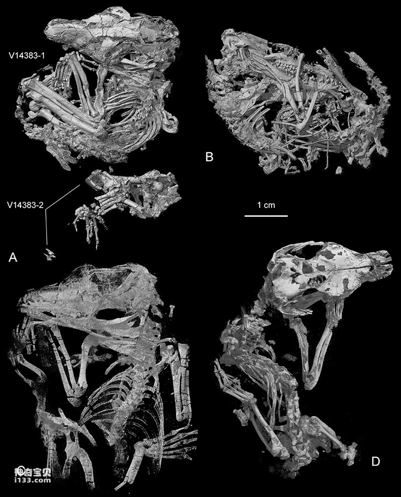
Figure 1. CT-reconstruction of Origolestes lii model (Photo provided by Mao Fangyuan)
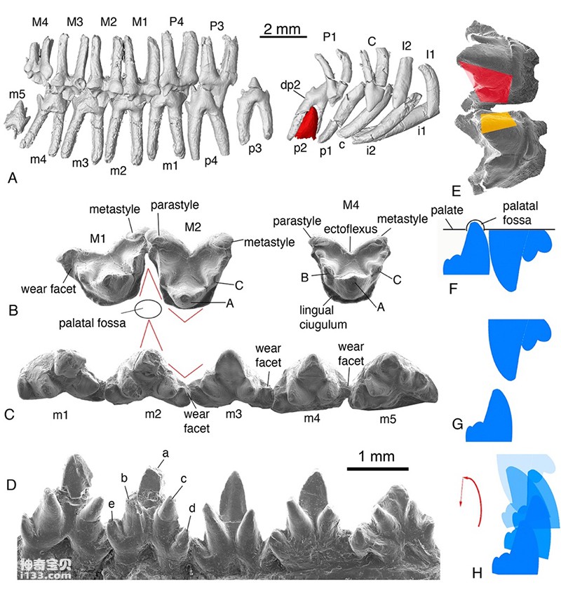
Figure 2. Origolestes lii’s tooth morphology, abrasion details and bite movement relationship (Photo provided by Mao Fangyuan)
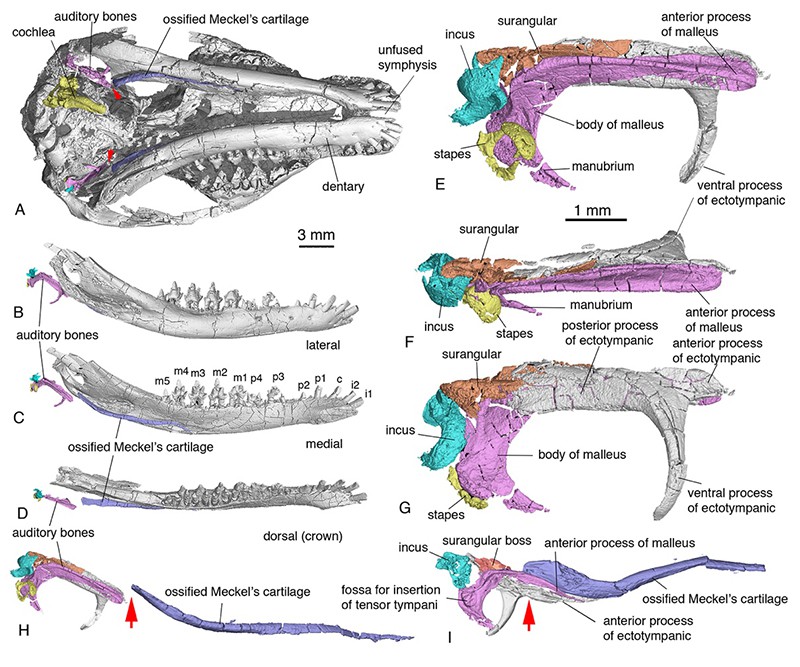
Figure 3. The morphology and anatomical position of the middle ear, inner ear, and Mckin's cartilage in Leygenus genus. H, Arrow points to the point where the ossicles separate from McMillon's cartilage. I, the arrow points to the bony connection between the transitional middle ear ossicles and Maxwell's cartilage in Acrodon (Photo provided by Mao Fangyuan)
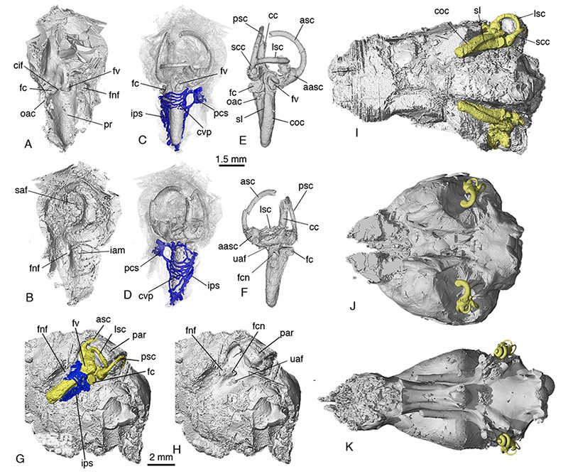
Figure 4. The structure of the inner ear, the brain cavity morphology and the relative relationship between E. leechei (A-I), and comparison with monotremes (J) and marsupials (K). Yellow is the inner mold of the inner ear structure, and blue is the related vascular system of the inner ear (Photo provided by Mao Fangyuan)
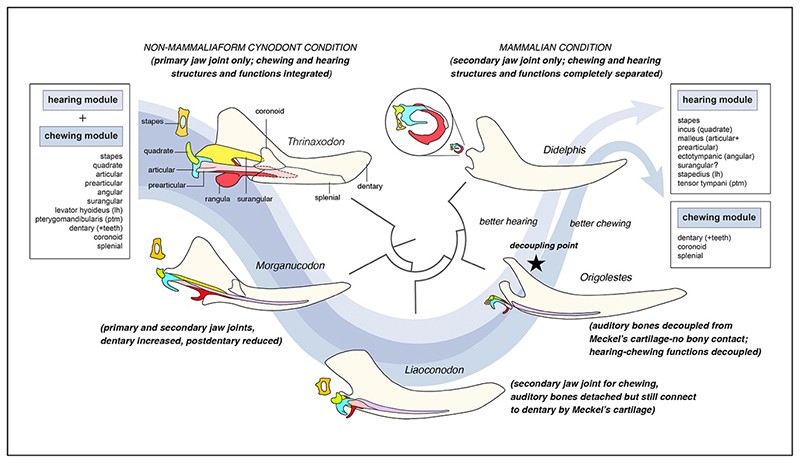
Figure 5. Schematic diagram of the modular evolutionary separation of hearing and chewing morphological functions in mammals (mammals) (Photo provided by Mao Fangyuan)
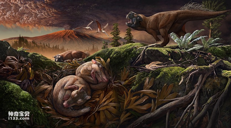
Figure 6. Ecological environment of Li's source predator and reconstruction of Lujiatun fauna (drawn by Zhao Chuang)
animal tags:
We created this article in conjunction with AI technology, then made sure it was fact-checked and edited by a Animals Top editor.
you may also like

Humanity's understanding of its own development history began with modern Homo sapiens, then early Homo sapiens, and then Homo erectus, and finally determined the status of Homo habilis and Australopithecus ape as human ancestors. Cro-Magnon This unde...

Among the large bears of the Miocene-Pliocene Epoch, the most famous are the Indian bear Indarctos and the suburban bear Agriotherium. Both species are large in size, widely distributed, and have a long history of discovery. They are representative species from the Miocene to Pliocene. Althou...

Humans are not always good at self-control, especially when the things they face seem both rich and mouth-watering. Although species extinctions often have multiple causes, the extinction of some species can be almost directly linked to the voracious appetites of modern humans. Read on to learn abou...

Algae are the lower types of the plant kingdom. There are many kinds of them, with an estimated 18,000 species, belonging to green algae, diatoms, golden algae, red algae and brown algae. They are widely distributed in various waters on the earth. Algae have a cell wall c...

When the American paleobotanist Letariak was analyzing and studying the Late Ordovician ancient soil in Pennsylvania, he discovered the fossilized footprints of some annelids or arthropods that entered deep into the soil from the surface. Based on these fossils, it is inferred that the terres...

The Tasmanian tiger is to Australia what the Sasquatch is to North America—a creature often seen by deluded amateurs but never actually seen in captivity. The difference, of course, is that Sasquatch is a complete myth, while the Tasmanian tiger is a real marsupial that only became extinct about a...

Reptiles are the earliest amniotes to appear on earth. They evolved from labyrinths among amphibians. The transition from amphibians to reptiles occurred in the Carboniferous Period, and the transition must have been marked by the production of amniotic eggs. In addition to the most important...

The Bible and Animals: The Deep Connection between Faith and NatureAs the core classic of Christianity, the Bible not only tells about human faith, morality and behavioral norms, but also The Bible shows the rich diversity of God's natural world, especially the animal world. Animals in the Bible...
Email: jsset668#gmail.com (change # to @) Please indicate your purpose of visit! Guangdong ICP No. 2022053326 XML| map| Chinese
