The British magazine Nature published online the research results of Wang Haibing and Wang Yuanqing of the Institute of Vertebrate Paleontology and Paleoanthropology, Chinese Academy of Sciences, and Meng Jin of the American Museum of Natural History on the evolution of the middle ear of early mammals: The discovery was reported in Lingyuan, Liaoning Province. A new genus of polytuberculous odonts from the Early Cretaceous, Jeholbaatar Kielanae, proposes a new model of mammalian middle ear evolution.
The latest study published on the mammal fossil was found in The Cretaceous Jiufotang Formation under Changzigou, Lingyuan, Liaoning. This fossil is related to A Beipiao sturgeon fossil is preserved on the same rock slab. After a long period of careful indoor repair, data processing and comparative research, the research team determined that the fossil represented a new species of the polytuberculate Eodontidae, and named it Jeholtherium gallii, with the genus name taken from Jehol Jehol Biota, this is the first report of polytuberculous odont fossils from the Jiufotang Formation. The species' original name is dedicated to Polish paleontologist Zofia Kielan-Jaworowska.
In the evolutionary history of vertebrates, the evolution of the mammalian middle ear is usually considered a classic case of biological recapitulation: the mammalian middle ear has experienced the transition from the Mandibular Mammalian Middle Ear to the Transitional Mammalian Middle Ear. , to the three evolutionary stages of the typical mammalian middle ear (Definitive Mammalian Middle Ear). This makes related research one of the hot spots in the study of early mammal evolution. However, the timing and mechanism of the occurrence of different middle ear evolutionary stages in various mammal lineages have always been difficult to study.
The complete middle ear structure is preserved in the holotype specimen of Jeholtherium galeni, providing direct evidence for studying the evolution of the ear region of early mammals. This research work reveals the complete morphology of each bone segment of the multituberculate middle ear and the contact relationship between them, and is helpful for exploring the evolutionary events of the postdentary bone of mammals from the mandibular middle ear to the typical mammalian middle ear. Supplemented with substantial puzzle pieces. Based on this study, researchers have a clearer understanding of the evolution of the surangular in mammals. This study reveals for the first time that in early mammals, the superior bone changed from an independent bone to a state that gradually healed with the body of the malleus and became the posterolateral part of the malleus. The malleus and incus in the new specimen are in complete shape and basically retain their original joint state. The two have an overlapping (dorsal-ventral) contact relationship. The researchers further proposed that during the evolution of the mammalian middle ear, although the morphology of the middle ear bones changed greatly, the malleus-incus joint showed two modes: overlapping joint and saddle joint.
Another important breakthrough in this study is that the researchers proposed a new model of the evolution of the middle ear in early mammals based on morphological and phylogenetic analysis results. Regarding the evolutionary mechanism from the mandibular middle ear to the typical mammalian middle ear, "cranial expansion" and "negative allometry" are two common hypotheses. The "brain expansion" hypothesis suggests that the enlargement of the brain during the growth of mammals causes the middle ear to move backward and eventually detach from the lower jaw. The "negative allometry" hypothesis emphasizes that in the early stages of embryonic development, the bone shape of the middle ear is larger than that of the mandible, and the middle ear ossifies earlier; therefore, in the later stages of embryonic development, as the skull and mandible increase, the middle ear The ear bones eventually break away from the jaw. With the progress of fossil research (eutricodonts) and embryonic development of living animals (monotremes and marsupials), the support for these two hypotheses continues to weaken. Another view is that the presence of ossified McFarland's cartilage and the eventual detachment of the postdentary bone may be related to the function of the mandible.
In the latest published paper, based on the results of phylogenetic analysis, researchers put forward a new hypothesis on the evolutionary mechanism of mammalian middle ear evolution in xenotherans (multituberculates + thieves). Among Mesozoic mammals, xenotherians had evolved the typical mammalian middle ear at least in the Middle/Late Jurassic (approximately 160 million years ago); while during the same period, or even later in the Early Cretaceous, other known All mammal groups also retain a transitional middle ear. At the same time, the dentary-squamous jaw joints of allothers are unique in that the joints are relatively open and can support large forward and backward movements of the mandible. They are morphologically and functionally different from the hinged dentary-squamous jaw joints in mammals. obvious. Combining the currently known morphology of the xenotherian middle ear and the differentiation time of the dentary-squamous jaw joint, the researchers proposed that in the evolution of the mammalian middle ear, the malleus-incus joint (primitive jaw joint) and the dentary bone -The squamosal jaw joints (secondary jaw joints) have co-evolved, and the overlapping primitive jaw joints can reduce the spatial constraints of the middle ear bones. The researchers proposed that there is also an evolutionary stage of transitional middle ear in the alien beasts, but the duration of this stage in the alien beasts is likely to be shorter than that of all other mammal groups. The evolutionary mechanism is probably due to the unique characteristics of the alien beasts. The dentary-squamous jaw joint and its feeding method provided more significant selective pressure for the middle ear to separate from the lower jaw than other taxa, thus accelerating the evolution of the middle ear, resulting in alien animals at least 160 million years ago (earlier than other All mammal groups) evolved the typical mammalian middle ear.
The three-dimensional scanning of the fossils in this study was completed at the High-Precision CT Scanning Center of the Key Laboratory of Vertebrate Evolution and Human Origins of the Chinese Academy of Sciences. The "Plate Fossil CT" (160-Micro-Computed Laminography) was used to conduct high-precision scanning of the fossils.
This research was supported by the Strategic Priority Science and Technology Project of the Chinese Academy of Sciences (Category B), the Basic Science Center Project of the National Natural Science Foundation of China (Cratonic Destruction and Terrestrial Biological Evolution), the National Natural Science Youth Fund, and the National Key Experiment in Modern Paleontology and Stratigraphy Supported by the Chamber Open Fund.
Article link: https://www.nature.com/articles/s41586-019-1792-0
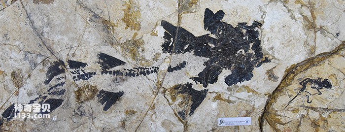
Figure 1. The holotype specimen of Rehe Junsei (lower right corner) and a Beipiao sturgeon specimen are preserved on the same rock slab (Photo courtesy of Wang Haibing)
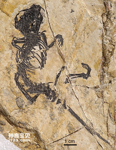
Figure 2. The holotype specimen of Jeholtherium gallbladders (Photo provided by Wang Haibing )
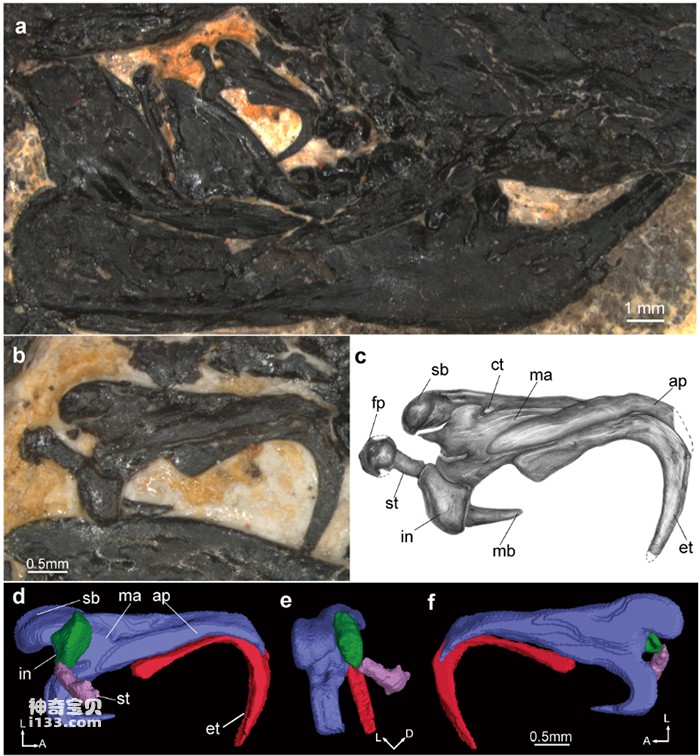
Figure 3. The left middle ear shape of the Jehol Jun beast (Photo courtesy of Wang Haibing)
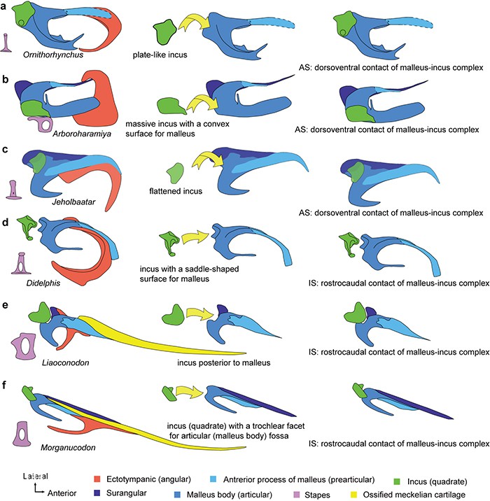
Figure 4. Two joint modes of malleus and incus in the evolution of the mammalian middle ear (Photo courtesy of Wang Haibing)
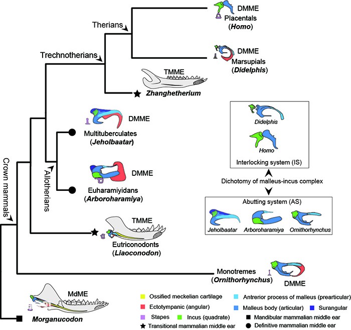
Figure 5. The evolutionary history of the middle ear of early mammals (Photo courtesy of Wang Haibing)

Figure 6. CL scan image of the holotype specimen of Jeholtherium gaiselii (Photo provided by Wang Haibing)
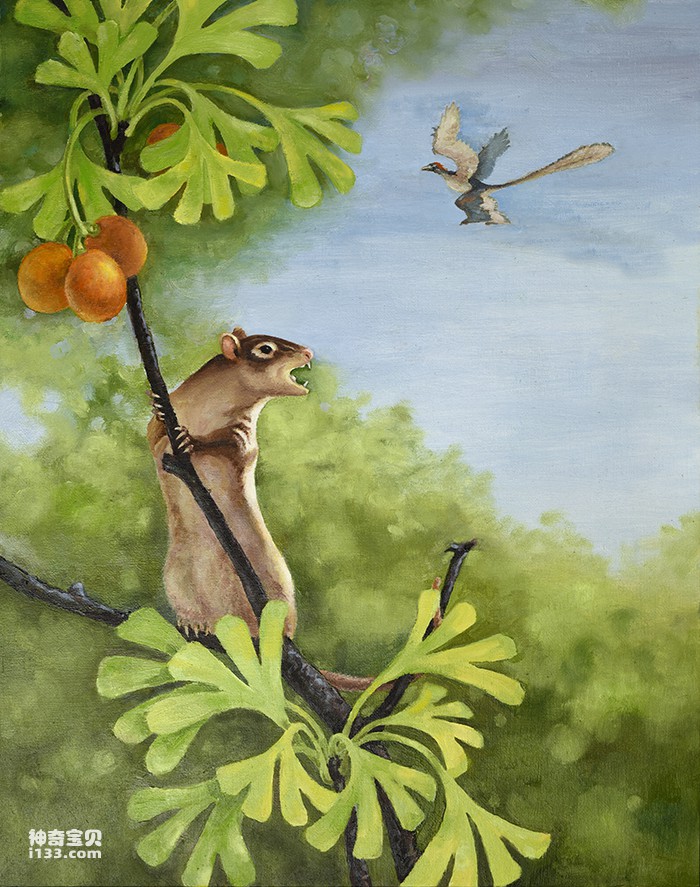
Figure 7. Rehabilitated Ecological Restoration Map of Junmao in Jehol (Photo provided by Xu Yong)
animal tags:
We created this article in conjunction with AI technology, then made sure it was fact-checked and edited by a Animals Top editor.
you may also like

Most conchovertebrates are small amphibians, adapted to life in shallow water and swamps. They first appeared in the Early Carboniferous Epoch and became extinct at the end of the Paleozoic Era, never flourishing. They are generally divided into three orders: Salamanders, Salamanders, and Amp...

When the ferns of the Paleozoic Era formed the first primeval forests on earth, gymnosperms, which were more advanced than ferns, had quietly appeared in the late Devonian period. But at that time, the climate on Earth was warm and humid, ferns developed more smoothly, and gymnosperms were no...

In order to understand and evaluate the phylogenetic position of the Tibetan woolly rhinoceros within the rhinoceros subfamily, Deng Tao et al. analyzed a group containing 17 species of Rhinoceros, including all 5 living species and 12 extinct species of rhinoceros. , including all four known...
")
Humans are the new generation of "Earth Overlords" after the dinosaurs, but the two appeared 58 million years apart. This is a long process, and many stories must have happened during this period. After the dinosaurs became extinct, what happened to the earth again? What did you experience? H...

Across the world, scientists have discovered a large number of tiny fossils from various marine strata from the Cambrian to the Triassic. Their shapes are very similar to various teeth, but they cannot be compared with any known animals. The teeth are so connected that scientists are still un...

作为能人的后代,在距今大约200万年前的非洲,直立人出现在人类进化的历史舞台上。直立人直立人的脑子已经明显增大,早期成员的脑量就已经达到800毫升左右,晚期成员则上升为1200毫升左右。而且,脑子不仅仅是体积增大了,它的结构也变得更加复杂并进行了重新改组,显示出直立人已经有了相当复杂的文...

In 1928, the famous American geological paleontologist Professor Gripp published a scientific masterpiece - "The Geological History of China". In this book, he proposed the term "Jehol Biota" for the first time to represent the comprehensive fossil group distributed in the wolf-finned fish ro...

As early as the late Triassic, when dinosaurs had just entered the evolutionary stage, a group of small animals that were inconspicuous at the time differentiated from therodonts among therapsid reptiles. They were born at the wrong time, because in the long years from the Jurassic to the Cre...
Email: jsset668#gmail.com (change # to @) Please indicate your purpose of visit! Guangdong ICP No. 2022053326 XML| map| Chinese
